Ryan P. Cabeen

Ryan P. Cabeen

Staff Machine Learning Engineer, Adjunct Assistant Professor of Research

ArteraAI

USC INI

Welcome! I am a computational imaging researcher and software developer dedicated to improving the capabilities and impact of biomedical imaging technology. I am currently a Staff Machine Learning Engineer at ArteraAI, where I am working on computer vision for AI-enabled predictive and prognostic cancer tests. Additionally, I am an Adjunct Assistant Professor of Research at the Stevens Neuroimaging and Informatics Institute (INI) at the University of Southern California (USC).

Prior to this, I conducted academic research in computer science and neuroscience at the USC Laboratory of Neuro Imaging with generous support from the Chan Zuckerberg Imaging Scientist program. I hold a PhD in Computer Science from Brown University, where I was advised by David H. Laidlaw, and a BSc from the California Institute of Technology. My research has centered on developing computational methods and software for characterizing the brains of humans and rodents using mathematical modeling, 3D visualization, and machine learning. Through interdisciplinary collaborations, we have applied these techniques to map normative changes throughout development and aging, as well as disruptions associated with dementia, environmental exposure, traumatic brain injury, and stroke.

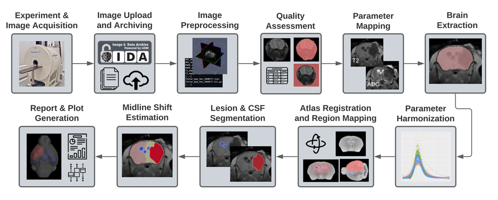
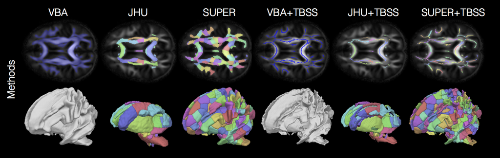
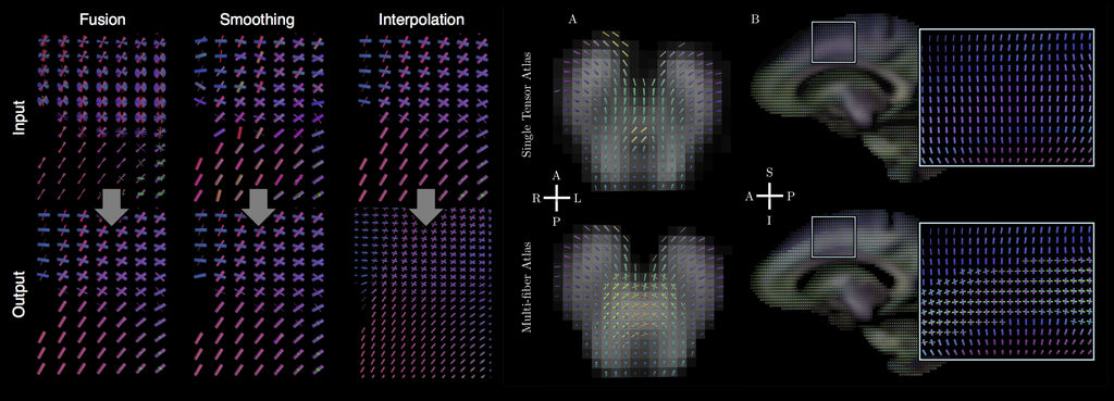
I invite you to learn more about my work by exploring my publications, some of which are listed below. Much of my research utilizes freely-available software that I developed, known as the Quantitative Imaging Toolkit (QIT). You can learn more about QIT by visiting its dedicated website.

Interests

  • Machine Learning
  • Biomedical imaging
  • Scientific Visualization
  • Computational Image Analysis
  • Software Development

Education

  • PhD in Computer Science, 2016

    Brown University

  • MSc in Computer Science, 2012

    Brown University

  • BSc in Eng. & Applied Science, 2005

    California Institute of Technology

Try the Quantitative Imaging Toolkit (QIT)

Software for 3D Visualization and Image Analysis. Automated and Interactive Tools for Diffusion MRI Tractography and Microstructure Imaging.

Learn more Download

Check out: a gallery of visualizations, a video of the atlas, and the abstract

Other Recent Publications

(2022). Anatomical and topographical variations in the distribution of brain metastases based on primary cancer origin and molecular subtypes: a systematic review. Neuro-Oncology Advances.

PDF DOI

(2022). Life after mild traumatic brain injury: Widespread structural brain changes associated with psychological distress revealed with multimodal magnetic resonance imaging. Biological Psychiatry Global Open Science.

PDF DOI

(2022). The Stroke Preclinical Assessment Network: Rationale, Design, Feasibility, and Stage 1 Results. Stroke.

PDF DOI

(2021). Connectivity characterization of the mouse basolateral amygdalar complex. Nature Communications.

PDF DOI

(2021). Microstructural properties within the amygdala and affiliated white matter tracts across adolescence. NeuroImage.

PDF DOI

(2021). Tractography dissection variability: what happens when 42 groups dissect 14 white matter bundles on the same dataset?. NeuroImage.

PDF DOI

(2020). Dietary Fructose Intake and Hippocampal Structure and Connectivity during Childhood. Nutrients.

(2020). Magnitude and timing of major white matter tract maturation from infancy through adolescence with NODDI. NeuroImage.

PDF

(2020). Tractography reproducibility challenge with empirical data (traced): The 2017 ismrm diffusion study group challenge. Journal of Magnetic Resonance Imaging.

(2019). Behavioral inhibition corresponds to white matter fiber bundle integrity in older adults. Brain imaging and behavior.

(2019). Clinical 7 T MRI: Are we there yet? A review about magnetic resonance imaging at ultra-high field. The British journal of radiology.

(2019). Harmonization of pipeline for preclinical multicenter MRI biomarker discovery in a rat model of post-traumatic epileptogenesis. Epilepsy research.

(2019). Image processing approaches to enhance perivascular space visibility and quantification using MRI. Scientific reports.

(2019). Limits to anatomical accuracy of diffusion tractography using modern approaches. NeuroImage.

(2019). MicroQIT - A Computational Framework for Population Microstructure Imaging. In Proceedings of the International Society for Magnetic Resonance in Medicine (ISMRM).

PDF

(2019). Nonparenchymal fluid is the source of increased mean diffusivity in preclinical Alzheimer's disease. Alzheimer’s & Dementia: Diagnosis, Assessment & Disease Monitoring.

(2019). Perivascular space fluid contributes to diffusion tensor imaging changes in white matter. NeuroImage.

(2019). Rapid and Accurate NODDI Parameter Estimation with the Spherical Mean Technique. In Proceedings of the International Society for Magnetic Resonance in Medicine (ISMRM).

PDF

(2018). Analytic tools for post-traumatic epileptogenesis biomarker search in multimodal dataset of an animal model and human patients. Frontiers in neuroinformatics.

(2018). Feasibility of Quantitative Diffusion MR Tractography of the Vestibulocochlear Nerve in Children with Unilateral Profound Sensorineural Hearing Loss. In Proceedings of the International Society for Magnetic Resonance in Medicine (ISMRM).

PDF